When comparing and contrasting a light microscope and an electron microscope, it’s essential to understand how they differ in principles, resolution, magnification, and applications. This guide provides a detailed light microscope and electron microscope comparison to help you choose the right tool for your scientific needs.

Electron microscopes, on the other hand, achieve higher resolution and magnification by using a beam of electrons. This allows electron microscopes to reveal details at the molecular and atomic levels, which light microscopes cannot. They are crucial for examining the ultrastructure of cells and materials.
The choice between these microscopes depends largely on your research needs. Light microscopes provide greater flexibility for biological studies, while electron microscopes are indispensable for high-resolution imaging. Understanding the strengths and limitations of each can help you determine the best tool for your scientific inquiries.

To compare and contrast light microscopes and electron microscopes, it’s vital to understand their underlying technologies. A light microscope uses visible light and optical lenses to magnify specimens, whereas an electron microscope employs a beam of electrons and electromagnetic lenses for superior resolution.
This light microscope and electron microscope comparison begins with their operational principles. Light microscopes use visible light passed through lenses to magnify the sample. Compound microscopes use multiple lenses to achieve higher magnification. Stereo microscopes provide three-dimensional views of specimens, ideal for dissection.
Electron microscopes use a beam of electrons instead of light. Transmission electron microscopes (TEMs) pass electrons through thin samples to view internal structures. Scanning electron microscopes (SEMs) scan the surface with electrons, creating detailed 3D images. These fundamental differences help us compare and contrast light microscope and electron microscope use in various scientific contexts.
Optical microscopes include compound and stereo microscopes.
Electron microscopes include TEMs and SEMs.
These fundamental differences define the specific uses and advantages of light and electron microscopes in various scientific fields.
 Light and electron microscopes differ significantly in magnification capabilities, resolution limits, and imaging techniques. Understanding these differences is crucial for selecting the appropriate tool for your scientific investigations.
| Feature | Light Microscope | Electron Microscope |
|---|---|---|
| Illumination | Visible Light | Electron Beam |
| Resolution | ~200 nm | ~0.1 nm |
| Magnification | Up to 1,000x | Up to 1,000,000x |
| Specimen Type | Living or Dead | Dead Only |
| Image Output | Color | Grayscale |
| Cost | Low | High |
| Sample Preparation | Minimal | Extensive |
A detailed comparison between light microscope and electron microscope reveals stark differences in magnification and resolution. While light microscopes can magnify up to 1,000x with ~200 nm resolution, electron microscopes can achieve up to 1,000,000x magnification and resolve structures as small as 0.1 nm.
Resolution also differs considerably. Light microscopes have a resolution limit of about 200 nanometers, constrained by the wavelength of light.
Electron microscopes, using electron beams, can resolve structures as small as 0.1 nanometers. Electrons have shorter wavelengths than photons, giving electron microscopes superior resolution.
Light microscopes use visible light as their illumination source. The image is formed on a glass slide and often viewed directly or through a camera. For better contrast, stains and dyes are commonly applied.
Electron microscopes use an electron beam instead of light. The electron beam interacts with the specimen, forming an image on a fluorescent screen or digital camera.
Since electrons can damage biological specimens, samples often require extensive preparation. This preparation includes coating with a thin layer of metal and operating in a vacuum environment to prevent scattering.
These distinctions, from illumination sources to image formation methods, highlight the unique applications and limitations of each type of microscope.
Electron microscopes provide significantly higher magnification and resolution—up to 1,000,000x and 0.1 nanometers, respectively. This makes them ideal for observing viruses, nanoparticles, and molecular structures that are invisible under a light microscope. While more expensive and complex, their capabilities are unmatched in fields like virology and nanotechnology.

In this light microscope and electron microscope comparison, it’s clear that each tool has specific strengths suited for different scientific fields. To properly compare and contrast light microscope and electron microscope, understanding their real-world use cases is key.
Light and electron microscopes play a crucial role in scientific advancements, with applications spanning various fields of research:
Microbiology & Pathology:
Light microscopes are widely used in microbiology for observing bacteria, fungi, and cell structures. Pathologists rely on them for histological studies, diagnosing diseases based on tissue samples.
Virology & Molecular Biology:
Electron microscopes are indispensable in virology, as they allow researchers to visualize viruses that are too small for light microscopes. For instance, electron microscopy played a pivotal role in imaging and understanding the structure of the SARS-CoV-2 virus during the COVID-19 pandemic. Learn more about its impact in virology in our dedicated post: The Role of Electron Microscopy in Virology: Why Seeing Is Believing.
Neuroscience & Cellular Biology:
Advanced imaging techniques such as Transmission Electron Microscopy (TEM) enable neuroscientists to study the ultrastructure of neurons and synaptic connections. This is critical in understanding neurodegenerative diseases like Alzheimer’s.
Materials Science & Nanotechnology:
In materials research, Scanning Electron Microscopes (SEM) are used to study surface morphology and microstructures of metals, polymers, and composites. TEM is also instrumental in nanotechnology for imaging nanoparticles and atomic structures.
By offering unique imaging capabilities, both light and electron microscopes contribute to breakthroughs in medicine, environmental science, and technological innovations.
In industrial settings and material sciences, light microscopes are commonly employed for quality control and inspection of materials. They are used to look at surface details, fractures, and crystalline structures. Their simplicity and cost-effectiveness make them a preferred option for many day-to-day applications.
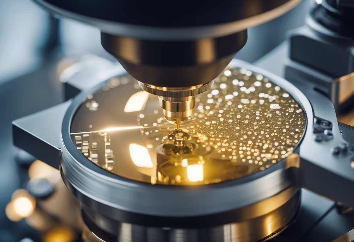
Electron microscopes are vital in materials science for analyzing the detailed composition of materials. They help in identifying defects in semiconductors, metals, and other industrial components. By providing detailed images at a nanometer scale, electron microscopes are indispensable in developing and improving new materials and technologies.

To compare and contrast light microscope and electron microscope effectively, we must evaluate not only their technical specifications but also their accessibility, costs, and training requirements.
Light microscopes are generally more affordable and accessible. They are widely used in education and routine laboratory work due to their low purchase and maintenance costs. These microscopes allow for the examination of living specimens, offering the added benefit of studying biological processes in real time.
Electron microscopes, on the other hand, are significantly more expensive. The initial purchase price is high, and ongoing maintenance costs can be considerable. Specialized training is required to operate these instruments, limiting their accessibility. These microscopes generally require complex preparations involving heavy metals and can only analyze dead specimens.
| Type of Microscope | Cost | Accessibility | Maintenance Cost | Specimen Type |
| Light Microscope | Low | High | Low | Living/Dead |
| Electron Microscope | High | Limited | High | Dead Only |
In terms of technical capabilities, light microscopes provide images in color, a significant advantage when differentiating between various cell components. However, they have low resolving power (around 200 nm) due to the longer wavelength of visible light, resulting in lower resolution images.
Electron microscopes excel in achieving high resolution (up to 0.1 nm), offering detailed images at the cellular and molecular levels. However, these images are typically in grayscale because electrons have a much shorter wavelength than visible light. The use of electron beams carries potential radiation risks, with the need for careful handling to avoid radiation leakage.
Training requirements for electron microscopes are also more stringent, necessitating advanced knowledge to handle these complex instruments. In contrast, light microscopes are user-friendly and require minimal training, particularly beneficial in educational settings.
| Type of Microscope | Resolution | Color/Grayscale | Radiation Risk | Training Needed |
| Light Microscope | Low (200 nm) | Color | None | Minimal |
| Electron Microscope | High (0.1 nm) | Grayscale | Present | Extensive |
| Type of Microscope | Advantages | Disadvantages |
|---|---|---|
| Light Microscope | – Affordable and widely accessible – Allows real-time observation of live specimens – Simple to use and requires minimal training – Provides color images of samples |
– Limited resolution (around 200 nm) – Maximum magnification of ~1,000x – Cannot reveal molecular or atomic structures |
| Electron Microscope | – Extremely high magnification (up to 1,000,000x) – Superior resolution (as small as 0.1 nm) – Provides detailed images of cellular and molecular structures – Essential for nanotechnology and materials science |
– Expensive to purchase and maintain – Requires extensive sample preparation – Can only be used on dead specimens – Requires specialized training to operate |
 In the realm of advanced microscopy techniques, you’ll encounter various methods like Near-Field Scanning Optical Microscopy (NSOM), Atomic Force Microscopy (AFM), and Scanning Electron Microscopy (SEM). These tools offer unique capabilities tailored to different scientific needs.
Near-Field Scanning Optical Microscopy (NSOM) breaks the diffraction limit of light using near-field techniques. The NSOM employs a sharp tip, usually coated with metal, brought extremely close to the sample. By scanning this tip in close proximity, NSOM achieves resolutions much finer than conventional light microscopy.
NSOM effectively combines optical and scanning probe techniques to provide high-resolution imaging. It is particularly useful for studying surfaces at the nanometer scale, offering detailed data about the optical properties of the nanoscale features. The method’s capability to achieve high spatial resolution makes it essential for materials science and biological research.
Atomic Force Microscopy (AFM) uses a fine-tipped probe that scans the surface of a sample to produce high-resolution images. The tip of an AFM microscope interacts with the sample at the atomic level, measuring forces between the tip and the surface. These interactions help generate three-dimensional images of the surface.
AFM is versatile, providing insights into a sample’s mechanical properties, such as elasticity and hardness. It doesn’t require conductive samples, unlike electron microscopy, expanding its usability. AFM’s precision makes it invaluable for applications in nanotechnology, materials science, and biophysics.
A Scanning Electron Microscope (SEM) uses focused beams of electrons to scan the surface of materials. SEM produces images by detecting secondary electrons emitted from the surface when bombarded with these electrons. This creates highly detailed, magnified images.
SEM is renowned for its high resolution and depth of field. It’s extensively used in material science, biology, and engineering to analyze surface topography and composition. The ability to produce three-dimensional images of the sample surface helps you examine intricate structural details.
1. What are the main differences between light and electron microscopes?
Light microscopes use visible light and glass lenses to magnify specimens, making them suitable for biological samples and live cells. Electron microscopes use electron beams, achieving much higher resolution but requiring extensive sample preparation.
2. Can a light microscope see viruses?
No, most viruses are too small for light microscopes, as their resolution limit is around 200 nanometers. Viruses, which are typically between 20-300 nanometers in size, require electron microscopes for visualization.
3. What are the disadvantages of electron microscopes?
Electron microscopes are expensive, require complex sample preparation, and can only be used on dead specimens. They also need vacuum conditions and specialized training for operation.
4. What type of microscope is best for viewing live cells?
Light microscopes, particularly phase-contrast or fluorescence microscopes, are ideal for viewing live cells as they do not require destructive sample preparation.
5. Why do electron microscopes require a vacuum?
Electron beams can be scattered by air molecules, which would distort the image. Operating in a vacuum prevents interference, allowing for clearer, high-resolution imaging.
In conclusion, this light microscope and electron microscope comparison outlines the distinct advantages of each. Whether you’re analyzing living tissues or investigating molecular structures, knowing how to compare and contrast light microscope and electron microscope will guide your decision-making. Choose wisely based on resolution needs, sample type, and technical resources. If you need assistance in selecting the right microscope for your work, contact us today for expert guidance and support.