Viruses are some of the smallest yet most dangerous entities known to science. Ranging from 20 to 300 nanometers, their structures are invisible to light microscopes. Enter electron microscopy, a powerful imaging tool that has transformed virology by allowing researchers to observe, classify, and understand viruses at a molecular level. Whether you’re developing vaccines, diagnosing viral infections, or studying viral evolution, electron microscopy is often the key to unlocking critical insights.
 What Is Electron Microscopy and How Does It Work?
Unlike light microscopes that use photons, electron microscopes rely on electron beams to generate high-resolution images. This technique enables magnification levels up to 10 million times, making it ideal for observing viruses in remarkable detail.
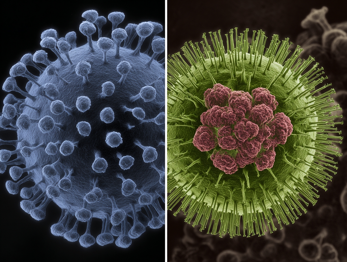
There are two main types:
Transmission Electron Microscopy (TEM): Produces detailed internal structures of viruses.
Scanning Electron Microscopy (SEM): Creates 3D surface images of viral particles.
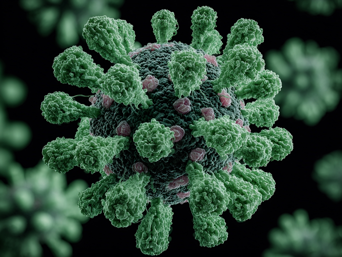
Electron microscopy allows direct visualization of virus particles, including their size, shape, and structural complexity. This is crucial for:
Virus classification
Studying morphology
Identifying new pathogens
One of the most common queries is: “When were viruses first seen under the electron microscope?”
The answer dates back to the 1930s, when the tobacco mosaic virus became the first virus observed using TEM. Today, EM remains a gold standard for early detection, especially in outbreak investigations.
Electron microscopy is often used when:
Routine diagnostic tests fail
Rapid identification is critical
Rare or emerging viruses are suspected
It’s particularly valuable in diagnostic pathology, where high-resolution imaging can confirm or rule out viral presence within tissue samples.
 Applications of Electron Microscopy in Virology
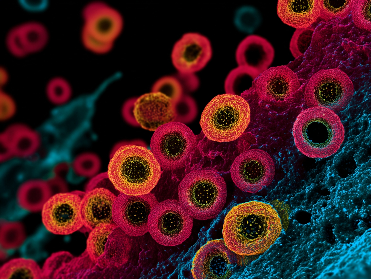
Detailed imaging helps researchers understand how viruses:
Attach to host cells
Inject genetic material
Replicate inside the host
Electron microscopy plays a key role in:
Visualizing antigenic structures
Designing effective vaccines (e.g., COVID-19 spike protein mapping)
Monitoring vaccine quality and stability
EM supports fundamental research in:
Viral replication cycles
Host-virus interactions
Antiviral drug mechanisms
Educational institutions often use electron micrographs to train new virologists and illustrate viral ultrastructure.
 Advantages of Electron Microscopy in Virology
High Resolution: Visualize viruses at the nanometer scale.
Detailed Morphology: Identify shape, size, and symmetry.
Rapid Diagnosis: Crucial for outbreak control.
Scientific Discovery: Enables observation of new viral strains.
Despite its power, electron microscopy does have drawbacks:
Expensive equipment
Requires skilled operators
Sample preparation is time-consuming
Can’t observe living viruses in real time
These factors mean EM is often used alongside other methods, like PCR or ELISA, rather than as a stand-alone diagnostic tool.
Looking for Electron Microscopes for Virology Research?
At Gold One Supplies, we work closely with trusted providers to help you source high-precision electron microscopes and accessories tailored to your lab’s needs. Contact us today to request a quote or learn more about available models and pricing.
 Frequently Asked Questions (FAQ)
Q1: What is the use of electron microscopy in virology?
It enables detailed visualization of viruses, aiding in diagnosis, classification, and vaccine development.
Q2: Which type of electron microscope is best for viruses?
Transmission Electron Microscopy (TEM) is typically used for internal structures, while Scanning Electron Microscopy (SEM) captures surface details.
Q3: Why can’t we use light microscopes to observe viruses?
Light microscopes lack the resolution needed to see objects smaller than ~200 nm. Most viruses are far smaller.
Q4: Can electron microscopy be used in disease diagnosis?
Yes, particularly for identifying unknown or rare viral infections when standard methods fail.
Q5: What are the disadvantages of using electron microscopy in virology?
Cost, complex operation, and the inability to observe live viruses are common limitations.